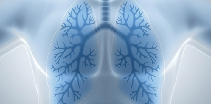
Bronchoscopy is a procedure to look directly at the airways in the lung called bronchus. Bronchoscope which is a thin flexible tube-like device having a very small camera at its tip is used for this procedure.
Why do we need bronchoscopy
- Unexplained symptoms related to chest, such as persistent cough, coughing up blood, wheezing and shortness of breath. The airways are examined for signs of problems and samples of tissue can be taken and examined for evidence of infection and cancer
- Persistent lung collapse is sometimes evaluated using bronchoscopy. This may reveal obstruction due to thick mucus ,tumor or foreign body. Obstruction can be removed and biopsies can be taken
- Radiological findings such as mass, nodules, infiltration can be evaluated by bronchoscopy; fluid samples or a biopsy are obtained to search for cancer, infection or other diseases.
- Various interventions (foreign body removal, bleeding control, electrocautery, cryotherapy, laser therapy, airway stent placement) can be administered via flexible bronchoscopy.
ENDOBRONCHIAL ULTRASOUND:
The diseases in the airways can be seen by bronchoscopy but sometimes diseases like cancer infection sarcoidosis can be outside of the airways.
Endobronchial ultrasound (EBUS) is a bronchoscopic technique that uses ultrasound along with bronchoscope to visualize lung tissue and mediastinum adjacent to airways .
Mediastinum is the part of the chest located between our lungs and contains the heart, windpipe, large blood vessels and lymph nodes . This area is hard to access without surgery; EBUS provides real-time imaging for the physician to easily view this areas and access lymph nodes for biopsy with the aspiration needle avoiding the need for surgical incision.

